FIGURE

Figure 5

ID
ZDB-FIG-240513-58
Publication
Song et al., 2024 - Functional and Genetic Analyses Unveil the Implication of CDC27 in Hemifacial Microsomia
Other Figures
All Figure Page
Back to All Figure Page
Figure 5

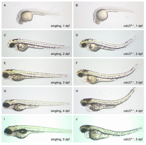
Phenotypic comparison between cdc27−/− mutants and wild-type zebrafish embryos from 1 to 5 days post-fertilization (dpf). This figure showcases the developmental progression and phenotypic variations observed in cdc27−/− mutant embryos compared to wild-type embryos from 1 dpf to 5 dpf. At 1 dpf (A,B), there are no significant phenotypic differences between the two groups. By 2 dpf (C,D), cdc27−/− embryos exhibit microcephaly, microphthalmia, and an abnormally curved spine, yet without noticeable abnormalities in the pharyngeal arch. At 3 dpf (E,F), the mutant embryos begin to show more pronounced phenotypic changes, including the absence of mandible structure formation, exacerbated spinal deformity, and the emergence of cardiac edema. These developmental anomalies, particularly the abnormal mandible development, persist in the mutant embryos at 4 dpf (G,H) and 5 dpf (I,J), highlighting the progressive nature of the craniofacial and skeletal deformities in cdc27−/− mutants.

Expression Data

Expression Detail
Antibody Labeling
Phenotype Data

Phenotype Detail
Acknowledgments
This image is the copyrighted work of the attributed author or publisher, and ZFIN has permission only to display this image to its users. Additional permissions should be obtained from the applicable author or publisher of the image. Full text @ Int. J. Mol. Sci.