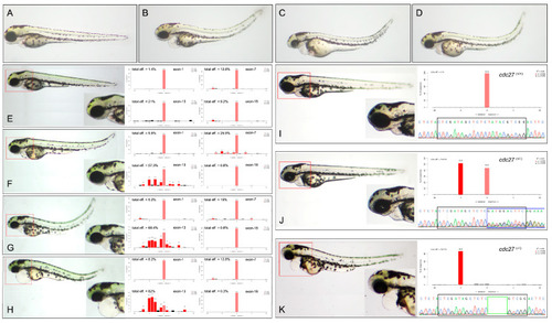
Comparison of phenotypes in wild-type and cdc27 gRNA injected zebrafish embryos. This figure presents the phenotypic differences observed in wild-type embryos (A) and those injected with cdc27 gRNAs (B–D) at 3 days post-fertilization (dpf). In over 90% of the gRNA injected embryos, a notable reduction in the overall size of craniofacial structures, along with spine malformation and cardiac edema, was evident compared to control siblings. Panels (E–H) illustrate the phenotypes and corresponding gene editing efficiency of embryos injected with four different gRNAs at 3 dpf. The severity of mandibular deformity in these embryos exhibited a progressive increase, with gRNA targeting exon-13 showing the highest gene editing efficiency. Moreover, a positive correlation was observed between the editing efficiency of gRNA (exon-13) and the severity of mandibular deformity. Panels (I–K) depict phenotypes, Sanger sequencing chromatograms, and genotypes of F2 embryos. The F2 generation exhibited two distinct phenotypic categories and three different genotypes: embryos without deformities had either wild-type or heterozygous (5 bp deletion) genotypes, while those with deformities were exclusively homozygous for the 5 bp deletion. The red box highlights enlarged zebrafish structures, the black box denotes the sequence of gRNA (exon-13) (GTC GAT AGC TCT CTA TAC GTC GG), the blue box represents Sanger sequencing bimodal patterns, and the green box indicates the 5 bp deletion sequence (TATAC).
|

