
Fig. 4
- ID
- ZDB-FIG-240509-23
- Publication
- Zhang et al., 2024 - ERK-activated CK-2 triggers blastema formation during appendage regeneration
- Other Figures
- All Figure Page
- Back to All Figure Page
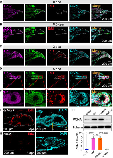
CK-2 promotes blastema formation by regulating cell proliferation. (A to D) Sections of regenerating legs stained with CK-2, p-ERK, and EdU at 0, 0.5, 3, and 5 dpa. The white dotted frame indicates the trochanter (0 dpa, 0.5 dpa) or the blastema (3 dpa, 5 dpa). Scale bars, 200 μm. (E) Micrograph was enlarged from (D) with 40×. Scale bar, 100 μm. (F and G) Detection of cell proliferation by EdU staining in response to CK-2 dsRNA injection. Nuclei were stained using DAPI (cyan). The white dotted frame indicates the blastema. Scale bars, 200 μm. (H) Detection of PCNA protein levels by Western blot. Uncut indicates uncut legs; am indicates 3-dpa regenerating legs; dsMock indicated 3-dpa regenerating legs were injected with Mock dsRNA, which was used as negative control. Each point represents the mean ± SD of three independent replicates. Student’s t test. |