
Fig. 1
- ID
- ZDB-FIG-240509-20
- Publication
- Zhang et al., 2024 - ERK-activated CK-2 triggers blastema formation during appendage regeneration
- Other Figures
- All Figure Page
- Back to All Figure Page
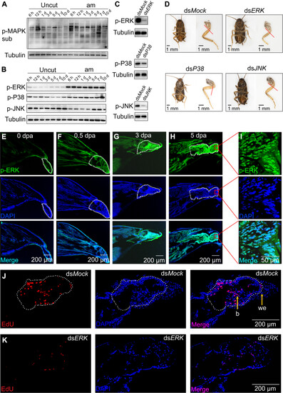
ERK was rapidly and sustained activated by phosphorylation to trigger blastema formation and leg regeneration. (A) Detection of MAPK pathway activity in the regenerating leg by Western blot. Uncut indicates uncut leg used as control; am indicates amputated leg; uncut and am also applies to the following legend. (B) Detection of p-ERK, p-P38, and p-JNK by Western blot. (C) In vivo double-stranded RNA (dsRNA) against ERK, P38, and JNK decreased the p-ERK, p-P38, and p-JNK levels, respectively. (D) ERK is required for leg regeneration. Injection of 1 μg of dsRNA against ERK, P38, and JNK and the regenerated legs were photographed. The red dashed line represents the amputation plane. Scale bars, 1 mm. (E to H) Immunofluorescence (IF) analysis of p-ERK in the regenerating leg after amputation. Nuclei were stained using DAPI (blue). The white dotted frame indicates the trochanter (0 dpa, 0.5 dpa) or the blastema (3 dpa, 5 dpa). Scale bar, 200 μm. (I) p-ERK was mainly localized in nuclei in the regenerating leg. Scale bar, 50 μm. (J and K) ERK is required for blastema formation during leg regeneration. Blastema formation was detected at 3 dpa by using 5-ethynyl-2-deoxyuridine (EdU) and 4′, 6-diamidino-2-phenylindole (DAPI) staining. The white dotted frame indicates the blastema. dsMock was used as a control. we, wound epidermis; b, blastema. Scale bars, 200 μm. |