
Fig. 3
- ID
- ZDB-FIG-240509-22
- Publication
- Zhang et al., 2024 - ERK-activated CK-2 triggers blastema formation during appendage regeneration
- Other Figures
- All Figure Page
- Back to All Figure Page
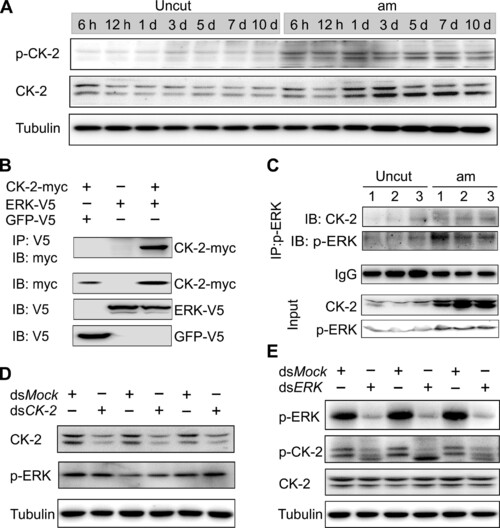
ERK phosphorylates and activates CK-2 to promote leg regeneration. (A) Patterns of p–CK-2 and CK-2 were up-regulated in the regenerating legs. (B) ERK combined with CK-2 by co-immunoprecipitation (co-IP). KC cells were cotransfected with the plasmids of ERK-V5 and CK-2–myc for 48 hours, and the cell extracts were immunoprecipitated (IP) with anti-V5 antibody, followed by immunoblot (IB) with anti-myc and anti-V5 antibody, respectively. (C) The combination between ERK and CK-2 was stronger in the regenerating legs than uncut group. The protein extracts (~1000 μg) of 3-dpa regenerating legs were immunoprecipitated with anti–p-ERK antibody, followed by IB with anti–CK-2 antibody or p-ERK antibody. Immunoglobulin G (IgG) indicated the antibody dose. (D) CK-2 has no effect on p-ERK. CK-2 dsRNA was injected for 48 hours. Protein levels of CK-2 and p-ERK were detected by Western blot using indicated antibody. Tubulin was used as loading control. Three biological replicates were performed. (E) ERK phosphorylates CK-2 in regenerating legs. ERK dsRNA was injected for 48 hours. Protein levels of p-ERK, p–CK-2, and CK-2 were detected by Western blot. Tubulin was used as loading control. Three biological replicates were performed. |