|
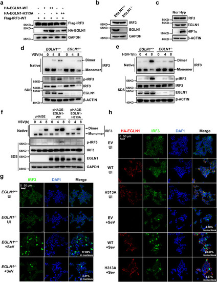
EGLN1 enhances IRF3 phosphorylation, dimerization, and nuclear translocation. a Immunoblotting of the indicated protein expression in HEK293T cells transfected with the plasmid expressing Flag-tagged IRF3 together with increasing amounts of HA-EGLN1 or HA-EGLN1-H313A. b Immunoblotting of the indicated protein expression in EGLN1+/+ or EGLN1−/− H1299 cells. c Immunoblotting of the indicated protein expression in H1299 cells under normoxia (Nor) (21% O2) and hypoxia (Hyp) (1% O2) for 4 h. dEGLN1+/+ and EGLN1−/− THP-1 cells were infected with VSV for the indicated times, and the cell lysates were analyzed by immunoblotting for monomeric (Monomer) and dimeric (Dimer) IRF3 (top; native-PAGE); phosphorylated IRF3 (p-IRF3), total IRF3, EGLN1, and β-ACTIN (bottom; SDS-PAGE). eEGLN1+/+ and EGLN1−/− THP-1 cells were infected with HSV-1 for the indicated times, and the cell lysates were analyzed by immunoblotting for monomeric (Monomer) and dimeric (Dimer) IRF3 (top; native-PAGE); phosphorylated IRF3 (p-IRF3), total IRF3, EGLN1, and β-ACTIN (bottom; SDS-PAGE). fEGLN1−/− H1299 cells stably expressed pHAGE empty vector, wild-type EGLN1 (WT) or the enzymatically inactive mutant (H313A) by lentivirus were infected with VSV for the indicated times, and the cell lysates were analyzed by immunoblotting for monomeric (monomer) and dimeric (dimer) IRF3 (top, native-PAGE); phosphorylated IRF3 (p-IRF3), total IRF3, EGLN1, and GAPDH (bottom; SDS-PAGE). gEGLN1+/+ or EGLN1−/− HEK293T cells were infected without (UI) or with SeV for 8 h and confocal microscopy image of endogenous IRF3 was detected by immunofluorescence staining using anti-IRF3 antibody. Scale bar = 50 µm. hEGLN1−/− HEK293T cells were transfected with the HA empty vector (EV), the plasmid expressing HA-tagged wild-type EGLN1 (WT) or the plasmid expressing the enzymatically inactive mutant (H313A), followed by infected without (UI) or with SeV for 8 h. Confocal microscopy image of endogenous IRF3 was detected by immunofluorescence staining using anti-IRF3 antibody and HA-tagged EGLN1 or its mutant (H313A) was detected by immunofluorescence staining with anti-HA antibody. Scale bar = 50 µm. Data in (a–h) are representative from three independent experiments. See also Supplementary Fig. 12. Source data are provided as a Source data file.
|