|
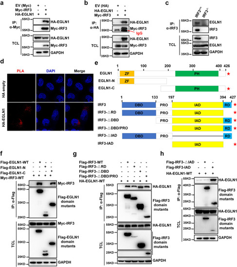
EGLN1 interacts with IRF3. a, b Co-immunoprecipitation of Myc-IRF3 with HA-EGLN1 and vice versa. HEK293T cells were co-transfected with the indicated plasmids for 24 h. Anti-Myc or anti-HA antibody conjugated agarose beads were used for immunoprecipitation and the interaction was detected by immunoblotting with the indicated antibodies. c Endogenous interaction between IRF3 and EGLN1 in IRF3-deficient (IRF3−/−) or wild-type H1299 cells (IRF3+/+). d In situ PLA assays of the EGLN1-IRF3 interaction in H1299 cells with the indicated combinations using anti-HA and anti-IRF3 antibodies, scale bar = 10 µm. e Schematic of EGLN1 domains interacting with IRF3 domains. The positive result of the interaction is indicated by the (★) signs. f Co-immunoprecipitation analysis of Myc-IRF3 with Flag-EGLN1 truncated mutants. HEK293T cells were co-transfected with the indicated plasmids. Anti-Flag antibody conjugated agarose beads were used for immunoprecipitation and the interaction was analyzed by immunoblotting with anti-Myc antibody. Flag-EGLN1 fragments (WT: full length; N, 1–196 aa; C, 130–426 aa). g, h Co-immunoprecipitation analysis of HA-EGLN1 with Flag-IRF3 truncated mutants. HEK293T cells were co-transfected with the indicated plasmids. Anti-Flag antibody conjugated agarose beads were used for immunoprecipitation and the interaction was analyzed by immunoblotting with anti-HA antibody. Flag-IRF3 fragments (WT: full length; ΔRD, 1–394 aa; ΔDBD, 133–427 aa; ΔDBD&PRO, 197–427 aa; ΔIAD, 1–197 & 394–427 aa; IAD, 197–394 aa). Flag-IRF3-ΔDBD&PRO expression was relatively lower compared to other fragments, its band was independently excised for longer exposure. EV empty vector, IP immunoprecipitation, TCL total cell lysates, PLA proximity ligation assay. Data in (a–d, f–h) are representative from three independent experiments. See also Supplementary Figs. 9 and 10. Source data are provided as a Source data file.
|