|
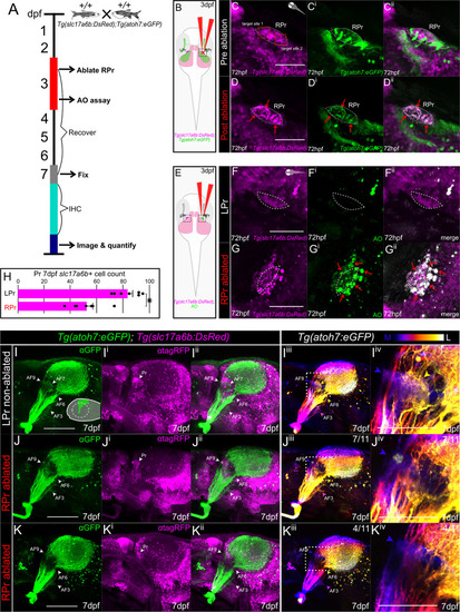
Tg(slc17a6b)-positive pretectal neurons are required for AF7 formation. (A) Experimental timeline: gsx1+/+;Tg(slc17a6b:DsRed);Tg(atoh7:eGFP) were raised until 72 hpf when they undergo unilateral ablation of the right pretectal (RPr) region. An acridine orange (AO) assay is performed on some samples at 72 hpf, hours post-ablation. Other ablated samples recover until 7 dpf. At 7 dpf samples are fixed and further undergo immunohistochemistry (IHC). (B) Schematic of unilateral RPr ablation in gsx1+/+;Tg(slc17a6b:DsRed);Tg(atoh7:eGFP) (magenta and green, respectively) at 72 hpf. LPr = left pretectum. (C-Cii) Pre-ablation of 72 hpf max projection of 2-photon z-stack through RPr separated by individual channels of Tg(atoh7:eGFP), green, Tg(slc17a6b:DsRed), magenta, and merged image channels. Two ablation sites are targeted and shown in C, red boxes. Dashed white outline indicates pretectal region. Orientation of sample is in right top corner. Scalebar = 50μm. (D-Dii) Post RPr ablation of same sample in C-Cii at 72 hpf, red arrows indicate displacement of the fluorescent proteins. (E) Schematic of imaging planes in black dashed boxes after unilateral RPr ablation and then following AO staining. (F-Gii) AO staining compared to intact LPr in the same RPr ablated sample, using Tg(slc17a6b:DsRed) (magenta) while red arrowheads indicate increased acridine orange (green) in ablated side. (H) Bar graph of quantified slc17a6b-positive neurons at 7 dpf in the LPr and RPr following 72 hpf RPr ablation. The ablated RPr showed statistically significant decreases in slc17a6b-positive neurons compared to LPr side (n = 7), t(13) = 0.96, p<0.001. (I-Kiv) Lateral 7 dpf max projections of confocal z-stacks of RGC axons in Tg(atoh7:eGFP) and Tg(slc17a6b:DsRed) and the same images depth color coded from medial (blue) to lateral (white), providing AF visualization, blue = AF9, yellow = AF7. Scalebar = 100μm. (I-Iiv) Control (no ablation, n = 6), orientation schematic in right bottom corner. (J-Jiv) 72 hpf unilateral RPr ablated resulting in partial AF7 disruption at 7 dpf (n = 7/11). (K-Kiv) 72 hpf unilateral RPr ablated resulting in total AF7 disruption at 7 dpf (n = 4/11). (Iiv, Jiv, Kiv) Zoomed in image from adjacent images with white dashed boxes for visualization of RGC axon AF patterns. Scalebar = 50μm.
|