
Fig. 7
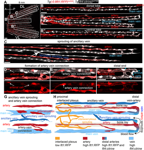
The fin vascular network expands through vein-derived sprouting of new veins and arteries. (A) Maximum intensity projections of confocal z-stacks of Tg(-0.8flt1:RFP)hu5333; Tg(flt4: citrine)hu7135 double-transgenic fish at 3 wpf (6 mm standard length) labelling arterial ECs (red) and venous ECs (white) in lateral views; anterior towards the left. Scale bar: 20 μm. (B) Dorsal lobe. (B′) Mid lobe. (B″) Ventral lobe. Scale bar: 30 μm. (C) Growth of ancillary vein. Scale bar: 30 μm. (D,E) Formation of artery-vein connections. Activation of arterial marker in single venous ECs (white arrows). Scale bar: 10 μm. (F) Distal end of caudal fin blood vessels. ECs transitioning from venous (blue arrowheads) to arterial (red arrowheads). Scale bar: 10 μm. (G) Schematic representation of ancillary vein sprouting and artery-vein connections. (H) Schematic representation of caudal fin vasculature at 3 wpf (6 mm standard length). |