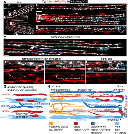
Fig. 7 The fin vascular network expands through vein-derived sprouting of new veins and arteries. (A) Maximum intensity projections of confocal z-stacks of Tg(-0.8flt1:RFP)hu5333; Tg(flt4: citrine)hu7135 double-transgenic fish at 3 wpf (6 mm standard length) labelling arterial ECs (red) and venous ECs (white) in lateral views; anterior towards the left. Scale bar: 20 μm. (B) Dorsal lobe. (B′) Mid lobe. (B″) Ventral lobe. Scale bar: 30 μm. (C) Growth of ancillary vein. Scale bar: 30 μm. (D,E) Formation of artery-vein connections. Activation of arterial marker in single venous ECs (white arrows). Scale bar: 10 μm. (F) Distal end of caudal fin blood vessels. ECs transitioning from venous (blue arrowheads) to arterial (red arrowheads). Scale bar: 10 μm. (G) Schematic representation of ancillary vein sprouting and artery-vein connections. (H) Schematic representation of caudal fin vasculature at 3 wpf (6 mm standard length).
Image
Figure Caption
Acknowledgments
This image is the copyrighted work of the attributed author or publisher, and
ZFIN has permission only to display this image to its users.
Additional permissions should be obtained from the applicable author or publisher of the image.
Full text @ Development

