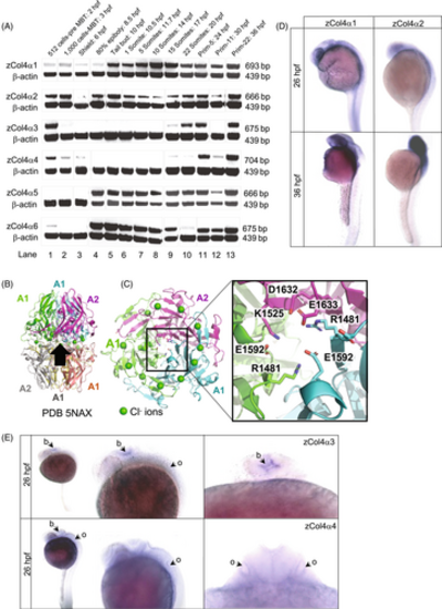
Embryonic expression of the six collagen IV α chains in zebrafish. (A) RT-PCR products for the zebrafish type IV collagen α chains at the indicated developmental stages (top band) range in size from 666 to 704 bp. The β-actin control band is shown below. Three expression patterns are noted: developmentally unrestricted (α1 and α2 chains); postgastrulation (α5 and α6 chains); and late-onset expression (α3 and α4 chains). Expression before 6 h postfertilization (hpf) stage is maternal. (B) Cartoon schematic of NC1 heterohexamer of α1 and α2 NC1 chains (PDB 5NAX). Black arrow shows viewpoint for C. (C) Bottom-up view of the heterotrimer. The inset is a close-up view of salt bridges that help form the heterotrimer interface. In the α1 and α2 chains, a glutamate (E1592 in α1 and E1633 in α2) forms a salt bridge with a basic residue (R1481 in α1 and K1525 in α2). (D) In situ hybridization for zebrafish α1 and α2 chains at 26 hpf and 36 hpf. (E) In situ hybridization for zebrafish α3 and α4 chains at 26 hpf, dorsal and lateral views. The α3 and α4 chain are localized in the developing brain (b), with α4 chain also detected in the otic vesicle (o). hpf, hours postfertilization, MBT, mid-blastula transition.
|
