|
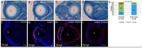
Erlotinib inhibits cell mass formation and phosphorylation of ERK1/2 in the ALE of plod3 mutants: (A–D) Histological sections from eyes of 4 dpf normal (A,B) or plod3 mutant (C,D) larvae. Larvae in (A,C) were treated from 1 to 4 dpf with vehicle (DMSO), whereas larvae in (B,D) were treated with Erlotinib. Scale bar is 20 µm. (E) Statistical analysis of the rescue of plod3 lens phenotype by Erlotinib as determined by localization of the lens. Outside, normal and inside represent localization of the lens relative to the retina as compared to the localization in normal larvae. (F–I) Single confocal sections from eyes of 75–76 hpf normal (F) or plod3 (G–I) mutant larvae, labeled with an antibody against pERK1/2 (red). Embryo in (H) was treated with DMSO and in (I) with Erlotinib. Nuclei (Dapi) are blue. L, lens. Scale bars are 20 µm.
|