|
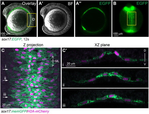
The morphology of gut endoderm at 12 s. (A-B) Images of a live embryo in which the endoderm is labeled with EGFP. (A-A″) Lateral view. (A) Overlay image of brightfield (A′) and epifluorescence image of EGFP (A″). (B) Dorsal view. Epifluorescence image of EGFP. (C-C′) Confocal images taken from a flat-mounted sample at a similar region outline in B (highlighted by yellow rectangles), showing endodermal cells labelled with the plasma membrane (GFP) and nuclei (pseudo-colored magenta). (C) Z projection of XY view. (C′) Images of XZ planes taken at the positions marked by i-iii in C. A, anterior; P, posterior; ML, mediolateral; D, dorsal; V, ventral.
|