|
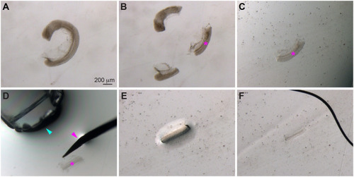
Deyolked embryonic tissue mounted on bridged slides. Bright-field images showing deyolked embryonic tissue mounted on a bridged coverslip. (A) A deyolked embryo. (B) An embryo was trimmed to obtain the desired tissue (magenta asterisk, used for imaging the gut endoderm). (C) The desired tissue was transferred in PBST to a 24×60 mm bridged coverslip. (D) PBST was removed by a glass pipette (cyan arrowhead), while the tissue was stabilized with a metal probe (magenta arrowhead). (E) The tissue after removal of PBST. (F) The tissue was mounted between the bridged coverslip and a 24×24 mm coverslip, and mounting medium was added, submerging the sample from the bottom left to the top right.
|