|
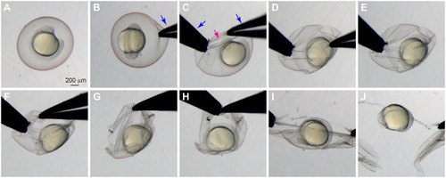
Manual dechorionation of live zebrafish embryos. Bright-field images illustrating the dechorionation process. (A) A live embryo before dechorionation. (B) A pair of forceps is used to gently pinch the chorion. (C) A second pair of forceps is used to pinch the chorion near the first pinch and to gently pull it apart. (D) The hole is made bigger, but not so much that the chorion collapses inward and squeezes the embryo. (E,F) Forceps are repositioned as needed to avoid squeezing the embryo. (G,H) Repositioning multiple times may be necessary. (I) Once the hole is large enough, the chorion is pulled apart without it collapsing inward and squeezing the embryo. (J) The embryo after dechorionation. Magenta arrow, hole in the chorion; blue arrows, forceps.
|