
FIGURE 11
- ID
- ZDB-FIG-230504-25
- Publication
- Bise et al., 2023 - The regeneration-responsive element careg monitors activation of Müller glia after MNU-induced damage of photoreceptors in the zebrafish retina
- Other Figures
- All Figure Page
- Back to All Figure Page
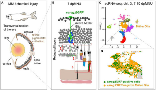
Identification of molecular differences between careg:EGFP-positive versus careg:EGFP-negative Müller glia during retina regeneration. (A) Schematic illustration of the experimental model in this study. (B) Cartoon of the retina at 7 dpMNU displays the induction of careg:EGFP expression in activated Müller glia (green cell), which give rise to the formation of proliferative progenitor cells (blue nuclei). The phenotypic defects of MNU-injury are highlighted in red frames: (1) Abolishment of actin filaments in synaptic photoreceptor processes in the outer plexiform layer. (2) Decrease of rod cell bodies in the outer nuclear layer. (3) Damage of inner segments of rod photoreceptors and the outer segments of UV-cones. (4) Distortion of photoreceptor outer segments. (C) UMAP visualization of cell clusters from the RNA-sequencing data of careg:EGFP retinas. (D) careg:EGFP-positive cells are detected mostly in the cluster of Müller glia. |