
FIGURE 1
- ID
- ZDB-FIG-230504-15
- Publication
- Bise et al., 2023 - The regeneration-responsive element careg monitors activation of Müller glia after MNU-induced damage of photoreceptors in the zebrafish retina
- Other Figures
- All Figure Page
- Back to All Figure Page
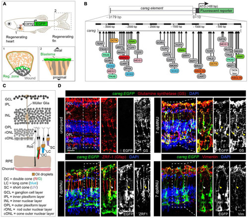
The careg regulatory element is activated in Müller glia during retina regeneration. (A) Schematic representation of the transgenic zebrafish line carrying the careg regulatory element upstream of the EGFP reporter, based on Pfefferli and Jaźwińska (2017). The careg:EGFP transgene is activated during heart (1) and fin (2) regeneration. (B) Prediction of transcription factor binding sites in the careg sequence with MatInspector (Genomatix) (Pfefferli and Jaźwińska, 2017). (C) Schematic representation of the retina in adult zebrafish, with an illustration of photoreceptors, based on Raymond and Barthel (2004) and Lagman et al. (2015). Outer segments of cones are colored according to their spectral sensitivity. Abbreviations are listed on legend at the bottom of the drawing. (D) Immunofluorescence stained sections of uninjured and 7 days post-MNU treatment (dpMNU) retinas of careg:EGFP (green) transgenic fish using three Müller glia markers (red): glutamine synthetase (GS), vimentin and the glial fibrillary acidic protein (GFAP, visualized with ZRF-1 antibodies). Nuclei are stained with DAPI (blue). In uninjured retina, no careg:EGFP is detected. At 7 dpMNU, careg:EGFP partially overlaps with some cells expressing GS, GFAP, and vimentin (arrowheads). In this, and all subsequent figures, a dashed frame demarcates the magnified area shown in adjacent panels. N ≥ 3 (number of fish). |