
FIGURE 5
- ID
- ZDB-FIG-230504-19
- Publication
- Bise et al., 2023 - The regeneration-responsive element careg monitors activation of Müller glia after MNU-induced damage of photoreceptors in the zebrafish retina
- Other Figures
- All Figure Page
- Back to All Figure Page
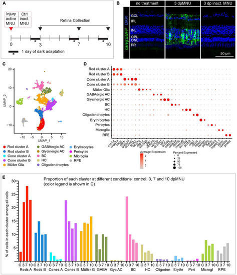
scRNA sequencing of careg:EGFP regenerating adult zebrafish retinas following MNU chemical injury. (A) Experimental design of retina isolation used for scRNA-sequencing. (B) Transversal sections of uninjured and regenerating careg:EGFP retinas at 3 days after treatment with harmful or inactivated MNU. careg:EGFP expression (green) is not detected after treatment with inactivated MNU, suggesting absence of injury. N = 3. (C) UMAP plots of the integrated cell RNA-sequencing data showing cluster assignments for each cell type collected from control and post-MNU treated retinas. (D) Dot plot showing expression of canonical markers for all retina cell types. Dot size indicates the proportion of cells expressing the corresponding gene and the color gradient indicates the average expression levels. (E) Histogram displaying percentage of cells in each cluster per time-point. |