
FIGURE 10
- ID
- ZDB-FIG-230504-24
- Publication
- Bise et al., 2023 - The regeneration-responsive element careg monitors activation of Müller glia after MNU-induced damage of photoreceptors in the zebrafish retina
- Other Figures
- All Figure Page
- Back to All Figure Page
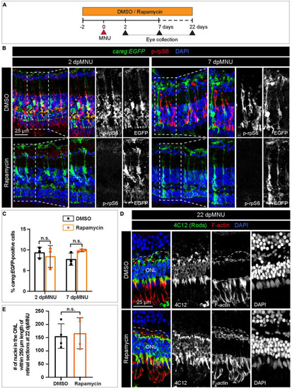
The careg element is not regulated by TOR signaling in regenerating retina. (A) Experimental design with 0.1% DMSO and 1 μM Rapamycin treatment. (B) Transversal sections of regenerating careg:EGFP (green) retinas at 2 and 7 dpMNU treated with DMSO or Rapamycin immunostained for the phosphorylated ribosomal protein p-rpS6 (red). Rapamycin treatment suppresses p-rpS6 immunoreactivity without affecting careg:EGFP expression (green). (C) Quantification of careg:EGFP-positive cells show a non-significant change between DMSO and Rapamycin-treated samples. Error bars, SEM. P-value was determined by two-way ANOVA with Sidák multiple comparisons test. n.s., not significant; N = 3. (D) Immunostaining of retina at 22 dpMNU demonstrates restoration of 4C12-positive rods (green) and F-actin-positive synaptic processes of the outer plexiform layer (encompassed with a dashed line). The position of outer nuclear layer (ONL) is indicated. (E) Quantification of nuclei in the outer nuclear layer within 250 μm length of retinal sections at 22 dpMNU. Error bars, SEM. P-value was determined by unpaired two-tailed Student’s t-test. n.s., not significant; N = 4. |