
Fig. 3
- ID
- ZDB-FIG-230312-41
- Publication
- Zeng et al., 2022 - Hypoxia-Responsive Subtype Cells Differentiate Into Neurons in the Brain of Zebrafish Embryos Exposed to Hypoxic Stress
- Other Figures
- All Figure Page
- Back to All Figure Page
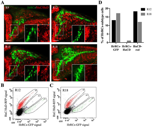
HrRCs in the brain did not express the early neuronal marker HuC/HuD. The images of fluorescence confocal microscopy were obtained from the hypoxia-exposure huORFZ embryos during recovery at different stages. (A) GFP-expressing HrRCs were observed under fluorescence microscopy at R9, R12, R15, and R18. The lower right two panels were amplified from the area indicated by the white dotted line box. (B–C) Data displayed the percentage of red-labeled HuC/HuD neurons overlapped with GFP-expressing HrRCs (at P8 gate) at R12 and R18 obtained through FACS sorting and immunostaining. (D) Calculation of the percentages of three different subtype cell populations isolated by FACS at R12 and R18. The scale bar is 20 µm. HrRCs: hypoxia-responsive recovering cells; FACS: fluorescence-activated cell sorting. |