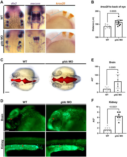
gldc deficient zebrafish display phenotypes consistent with Gldc deficient mammals. (A) WISH analysis of mecom in WT and gldc morphants revealed an altered rhombomere four. gldc morphants exhibited disorganized migrating neural crest cell populations marked with dlx2. krox20, a gene expressed in rhombomeres three and five, appeared shifted in comparison to the back of the eye. Scale bar = 50 μM. (B) Quantification of the distance between the edge of rhombomere three and the back of the eye. (C) Injections of dextran-rhodamine into the brain ventricles of live WT and gldc morphant animals at 24 hpf. In gldc morphants, brain ventricles appear enlarged. Scale bar = 50 μM. (D) Acridine Orange assay in WT and gldc morphants at 24 hpf; red arrowheads indicate example AO+ cells. Scale bar = 50 μM. (E,F) Quantification of AO+ cells in the brain and kidney, respectively. Distance measurements and cell counts were compared by unpaired T-tests. Significant differences are shown above the brackets.