
Figure 7
- ID
- ZDB-FIG-221226-260
- Publication
- Weaver et al., 2022 - gldc Is Essential for Renal Progenitor Patterning during Kidney Development
- Other Figures
- All Figure Page
- Back to All Figure Page
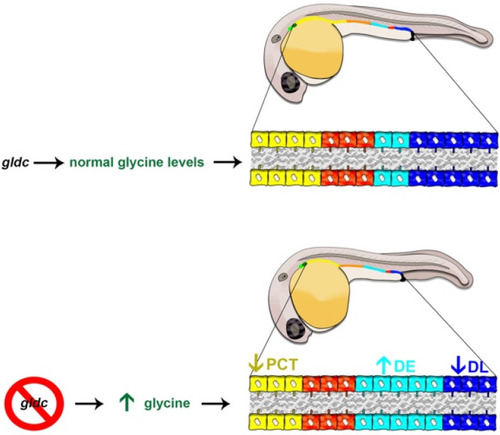
Nephrogenesis in the WT zebrafish embryo compared to gldc deficiency which leads to lethal elevated glycine levels. (Top) At 24 hpf, when the nephron segment pattern is established, WT embryos exhibit nephrons tubules with a series of four segments: the PCT (yellow-colored cells), PST (orange-colored cells), DE (turquoise-colored cells) and DL (dark blue colored cells). (Bottom) nephron tubule segment populations are altered in gldc deficient zebrafish, such that the PCT segment is decreased in size (arrow down), while the DE segment population is expanded (arrow up) and the DL segment is decreased (arrow down). These alterations correlate with changes in the expression of essential renal progenitor genes, observations that provide a crucial foundation for future studies to elucidate the underlying molecular mechanisms of these nephrogenesis defects. |