|
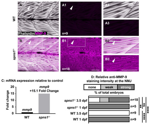
mmp9 mRNA and protein are upregulated in spns1−/− larvae at 3.5 dpf. (A,B) Anterior left, dorsal top, side-mounted larvae. Phalloidin (white) to visualize actin and MMP-9 antibody staining (pseudo-colored fuchsia). (A,A2,B,B2) Merged phalloidin and MMP-9 channels and (A1,B1,A3,B3) MMP-9 single channel. (A1) MMP-9 is not detected in WT muscle. (B1) MMP-9 is detected in spns1−/− muscle. White arrowheads highlight MMP-9 staining (or lack thereof in wild-type larvae). (C) Relative mmp9 mRNA expression in WT and spns1−/− larvae measured as fold change in 3.5 dpf larvae. (D) Percentage of spns1−/− and WT at 1 and 3.5 dpf with qualitatively scored relative fluorescence intensity. ** p < 0.01, **** p < 0.0001. Scale bars 50 µm.
|