|
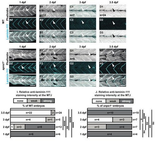
Normal initial muscle development is followed by abnormal 3.5 dpf laminin-111 levels in spns1−/− embryos and larvae. (A1–H3) Anterior left, dorsal top, side-mounted embryos and larvae stained with phalloidin (white) to visualize actin and laminin-111 antibodies (cyan). (A1–H1) Phalloidin staining. (A2–H2) Laminin-111 staining. (A3–H3) Merged phalloidin and laminin-111 channels. White arrowheads point to laminin-111 localized to the MTJ. Laminin-111 staining, not detected in WT embryos and larvae at 3–3.5 dpf, is detected in (H2–H3) spns1−/− 3.5 dpf larvae. (I,J) Relative fluorescent intensity of laminin-111 protein in WT and spns1−/− embryos and larvae over time (see methods). * p < 0.05, ** p < 0.01, **** p < 0.0001. Scale bars 50 µm.
|