FIGURE

Figure 6.

ID
ZDB-FIG-220814-6
Publication
Zhang et al., 2022 - A transgenic zebrafish for in vivo visualization of cilia
Other Figures
All Figure Page
Back to All Figure Page
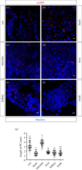
Figure 6.

Cilia in adult sx1001 tissue were uniformly labelled. (a–f) Transverse cryosection of different tissues to observe the distribution of sx1001 labelled (red) cilia in adult zebrafish (n = 3 for each sample). The nucleus was stained by Hoechst (blue). Scale bar, 5 μm. (a) Cilia at the photoreceptor cell layer; (b) cilia of myocardial cell cilia; (c) cilia structure of the small intestine; (d) cilia of liver cells; (e) cilia of renal cell; (f) cilia of skeletal muscles. (g) The length of tissue cilia was measured using the MIP method previously described. Each sample has been measured with the length of 40 straight primary cilia (n = 5 for each sample).

Expression Data

Expression Detail
Antibody Labeling
Phenotype Data

Phenotype Detail
Acknowledgments
This image is the copyrighted work of the attributed author or publisher, and ZFIN has permission only to display this image to its users. Additional permissions should be obtained from the applicable author or publisher of the image. Full text @ Open Biol.