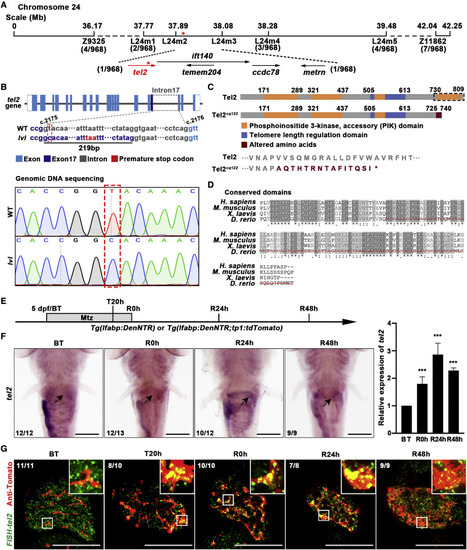
Figure 2. tel2 is the candidate gene of the lvl mutant (A) Genetic map of the candidate region on chromosome 24. Numbers below SSLP markers indicate recombination events. (B) Top panel: diagram showing the genomic structure of tel2. Bottom panel: sequencing result showing the DNA sequence change in lvl. A T-to-C transversion (red dashed box) was positioned in the pre-mRNA splice site of tel2 intron 17 (between coding DNA nucleotides 2,175 and 2,176), leading to the 219-bp insertion and introducing a premature stop codon in the coding region. (C) Top panel: schematic showing the difference of function domains (black dashed box) in Tel2 between the wild type (WT) and lvl. Bottom panel: the protein sequence change in the lvl mutant. (D) Zebrafish Tel2 and its homologous proteins in human, mouse, and Xenopus, showing a highly conserved phosphatidylinositol 3-kinase (PI3K) function domain. The red dashed line represents mutated amino acids in lvl. (E) Experimental schematic illustrating the stages of Mtz treatment and analysis at BT, T20h, R0h, R24h, and R48h. (F) Whole-mount in situ hybridization (WISH) images showing tel2 expression in the liver region (arrows) and quantitative real-time PCR showing the relative expression level of tel2 in regenerating livers from BT to R48h. n = 3 technical replicates. Values represent means ± SD. ∗∗∗p < 0.001 by t test. (G) Fluorescence in situ hybridization (FISH) and antibody staining in regenerating livers showing expression of tel2 (green) in Tp1+ cells (red). Expression of tel2 was upregulated from T20h and up to the maximal level at R24h. Numbers indicate the proportion of larvae exhibiting the expression shown. Scale bars, 100 μm (F and G). See also Figure S2.
|
