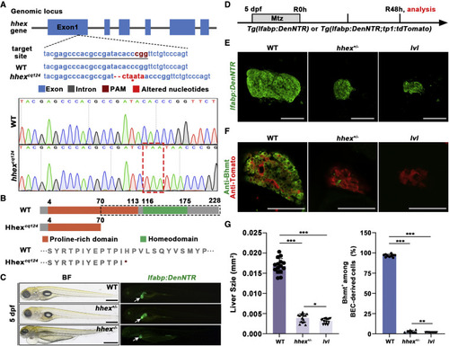
Figure 5. hhex+/− heterozygotes show liver regeneration defects similar to lvl (A) The genomic structure of hhex (top panel) and the guide RNA (gRNA) target sequence in hhex exon 1 (center panel). Bottom panel: sequence alignment showing the 2-bp deletion and 6-bp addition containing a premature stop codon (red asterisk/dashed box) in the hhexcq124 mutant. (B) Top panel: schematic showing the function domain change (black dashed box) of Hhex in the WT and hhexcq124 mutant. Bottom panel: the protein sequence change in the hhexcq124 mutant. (C) BF and epifluorescence images showing the body phenotypes of WT sibling, heterozygous mutant (+/−), and homozygous (−/−) mutant at 5 dpf (n = 25–27 larvae). hhex−/− mutants exhibit severe defects in multiple organs, including the liver, but hhex+/− mutants commonly survive without noticeable flaws. Arrows point to the liver. (D) Experimental schematic illustrating Mtz treatment and analysis at R48h. (E) Confocal projection images showing regenerating livers of WT, hhex+/−, and lvl at R48h. (F) Single optical section images showing Tomato (red) and Bhmt (green) expression in regenerating livers of WT, hhex+/−, and lvl at R48h. (G) Graph showing quantification of liver size shown in (E) (n = 15 larvae) and the percentage of Bhmt+ among BEC-derived cells shown in (F) (n = 10 larvae). Values represent means ± SD. ∗p < 0.05, ∗∗p < 0.01, ∗∗∗p < 0.001 by t test. Scale bars, 100 μm (E and F) and 400 μm (C). See also Figure S5.
|
