
Fig. 8
- ID
- ZDB-FIG-220801-250
- Publication
- Eugenin von Bernhardi et al., 2022 - A versatile transcription factor: Multiple roles of orthopedia a (otpa) beyond its restricted localization in dopaminergic systems of developing and adult zebrafish (Danio rerio) brains
- Other Figures
- All Figure Page
- Back to All Figure Page
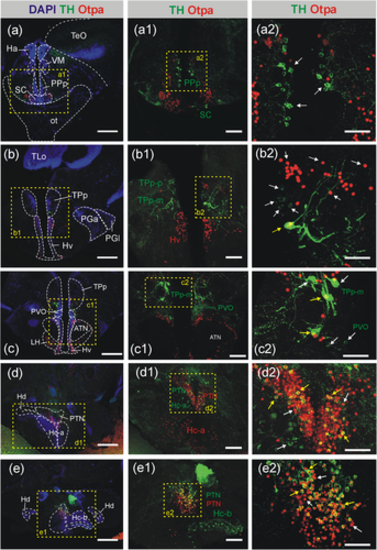
Confocal microscopic analysis of TH- and Otpa-positive cells in the adult zebrafish preoptic region and diencephalon at levels of the periventricular posterior tuberculum, posterior tuberal nucleus, and caudal periventricular hypothalamic zone. Note that a1/a2-e1/e2 are separate enlarged microphotographs of these transverse sections. The entire preoptic complex shows no cellular co-localization of TH and Otpa (a-b) as demonstrated for the posterior parvocellular preoptic (PPp) and suprachiasmatic nucleus (SC; a-a2; white arrows). Only the pear-shaped magnocellular part of the periventricular posterior tuberculum (TPp-m; b-b2/c-c2) shows cellular colocalization of TH and Otpa, but not the parvocellular nucleus of the periventricular posterior tuberculum (TPp-p), as demonstrated in magnifications (b1-c1/b2-c2, white and yellow arrows, respectively). Similarly, in the posterior tuberal nucleus (PTN) many cells show co-localization of TH and Otpa (d1-d2/e1-e2, white versus yellow arrows). Note complete absence of such overlap in the paraventricular organ (PVO; c1-c2) and the entire caudal hypothalamus (Hc; d1/e1), but TH immunostain in liquor-contacting cells of the ventral posterior recess lining of the Hc-b (e1). In both, TPp-m and PTN many–but not all—cells show colocalization of TH and Otpa. Scale bars = 250 µm (a-e), 100 µm (a1-e1), 50 µm (a2-e2). Abbreviations: ATN, anterior tuberal nucleus; Ha, habenula; Hc-a, caudal zone of periventricular hypothalamus in front of posterior recess; Hc-b, caudal zone of periventricular hypothalamus around posterior recess; Hd/Hv, dorsal/ventral zone of periventricular hypothalamus; LH, lateral hypothalamic nucleus; ot, optic tract; PGa/PGm, anterior/medial preglomerular nucleus; PPp, posterior parvocellular preoptic nucleus; PTN, posterior tuberal nucleus; PVO, paraventricular organ; SC, suprachiasmatic nucleus; TeO, optic tectum; TLo, torus longitudinalis; TPp, periventricular nucleus of posterior tuberculum; TPp-m, magnocellular cells of TPp; TPp-p, parvocellular cells of TPp; VM, ventromedial thalamic nucleus |