
Fig. 12
- ID
- ZDB-FIG-220801-254
- Publication
- Eugenin von Bernhardi et al., 2022 - A versatile transcription factor: Multiple roles of orthopedia a (otpa) beyond its restricted localization in dopaminergic systems of developing and adult zebrafish (Danio rerio) brains
- Other Figures
- All Figure Page
- Back to All Figure Page
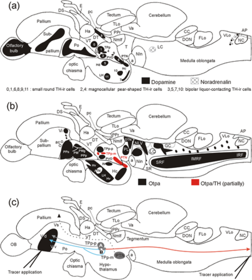
Schematic lateral view of adult zebrafish brain shows summary of (a) TH-positive brain nuclei (modified from Rink & Wullimann, 2002a; Wullimann & Rink, 2002), and (b) Otpa domains. (c) Schematic lateral view of adult zebrafish brain shows telencephalic and spinal connections of posterior tubercular nuclei. In (a), Arabic numbers, which were initially introduced in our developmental studies (Rink & Wullimann, 2002a; Wullimann & Rink, 2002), are also indicated for diencephalic and hypothalamic dopaminergic cell groups. Note that descending spinal projections of dopaminergic parvocellular periventricular posterior tubercular cells (TPp-p; Becker et al., 1997), and ascending projections of dopaminergic positive TPp-p and magnocellular pear-shaped periventricular posterior tubercular cells (TPp-m; Rink and Wullimann, 2001) have been previously established in the adult zebrafish brain (in larvae: Tay et al., 2011). Furthermore, TH-negative posterior tuberal (PTN) cells project to the zebrafish pallium (Rink & Wullimann, 2001, 2004; confirmed in goldfish by Northcutt, 2006). We propose the hypothesis that there are two populations of TPp-m cells, one with exclusive ascending projections to the striatum, and one with additional spinal projections, plus additional TPp-p projections to the striatum as well as ascending projections to the pallium from the posterior tuberal nucleus. (c) is modified from Rink and Wullimann (2002b). Abbreviations: A, ansulate commissure, ac anterior commissure; AP, area postrema; ATN, anterior tuberal nucleus; CC, crista cerebellaris; DON, descending octaval nucleus; DS, saccus dorsalis; DT, dorsal thalamus; DTN, dorsal tegmental nucleus; E, epiphysis; Flo, facial (sensory) lobe, GC, central gray; Ha, habenula; Hc/Hd/Hv, caudal/dorsal/ventral zone of periventricular hypothalamus; IN, intermediate hypothalamic nucleus; LC, locus coeruleus; LH, lateral hypothalamic nucleus; LR lateral hypothalamic recess; NC, commissural nucleus of Cajal; NLV, nucleus lateralis valvulae; Nmlf, nucleus of medial longitudinal fascicle; NIn, interpeduncular nucleus; OB, olfactory bulb; oc, optic chiasma; pc, posterior commissure; PL, perilemniscal nucleus; PM, magnocellular parvocellular preoptic nucleus; PMg, gigantocellular part of PM; Po, preoptic region; PPa/PPp, anterior/posterior parvocellular preoptic nucleus; PM, magnocellular preoptic nucleus; PPr, periventricular pretectum; PTN, posterior tuberal nucleus; PVO, paraventricular organ; SR, superior raphe; SRF, superior reticular formation; T, midbrain tegmentum; TLo, torus longitudinalis; TPp-m, magnocellular cells of the periventricular posterior tubercular nucleus; TPp-p, parvocellular cells of periventricular posterior tubercular nucleus; Va, valvula cerebelli; VLo, vagal (sensory) lobe; Vd/Vi//Vs/Vv, dorsal/intermediate/supracommissural/ventral nucleus of ventral telencephalon; VT, ventral thalamus |