
Fig. 6
- ID
- ZDB-FIG-220801-248
- Publication
- Eugenin von Bernhardi et al., 2022 - A versatile transcription factor: Multiple roles of orthopedia a (otpa) beyond its restricted localization in dopaminergic systems of developing and adult zebrafish (Danio rerio) brains
- Other Figures
- All Figure Page
- Back to All Figure Page
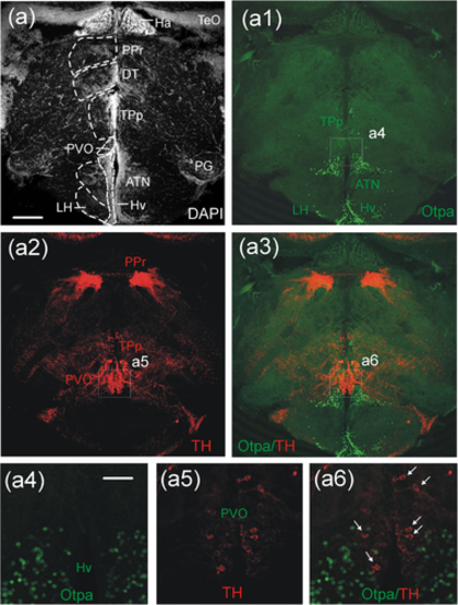
Confocal microscopic analysis of catecholaminergic (TH) and Otpa-positive cells in the adult zebrafish diencephalon at the level of the paraventricular organ. An ideal transverse section for displaying the periventricular pretectum (PPr), dorsal thalamus (DT), periventricular nucleus of the posterior tuberculum (TPp), ventral periventricular hypothalamic zone (Hv), as well as anterior tuberal and lateral hypothalamic nuclei (ATN/LH) is shown for DAPI stain (a), and Otpa (a1), TH (a2) and merged Otpa/TH immunostains (a3). Separate enlarged microphotographs show Otpa (a4), TH (a5) and merged Otpa/TH (a6) immunostained cells and demonstrate that no cellular colocalization occurs in the paraventricular organ (PVO; white arrows), as is obviously also the case for the periventricular nucleus of the posterior tuberculum (TPp), the periventricular pretectum (PPr), the ventral zone of the periventricular hypothalamus (Hv), and the anterior tuberal and lateral hypothalamic nuclei (ATN/LH). Scale bars = 250 µm (a), 125 µm (a4), applies also to (a5/a6). Abbreviations: ATN, anterior tuberal nucleus; DT ,dorsal thalamus; Ha, habenula; Hv, ventral zone of periventricular hypothalamus; LH, lateral hypothalamic nucleus; PG, preglomerular complex; PPr, periventricular pretectum; PVO, paraventricular organ; TeO, optic tectum; TPp, periventricular nucleus of posterior tuberculum |