
Fig. 4
- ID
- ZDB-FIG-220801-246
- Publication
- Eugenin von Bernhardi et al., 2022 - A versatile transcription factor: Multiple roles of orthopedia a (otpa) beyond its restricted localization in dopaminergic systems of developing and adult zebrafish (Danio rerio) brains
- Other Figures
- All Figure Page
- Back to All Figure Page
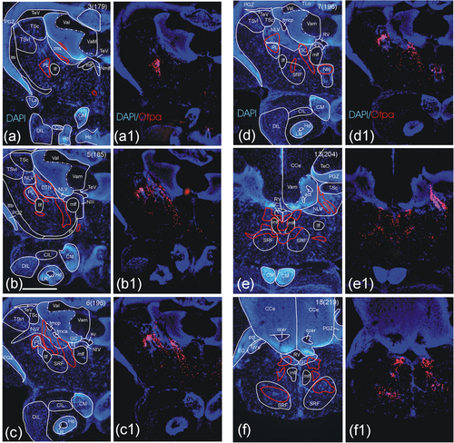
Distribution of Otpa-positive cells in the adult zebrafish midbrain and anterior hindbrain. Epifluorescent microscopic analysis shows Otpa domains in anterior midbrain and hindbrain regions. (a–f) A series of sequential transverse sections shows DAPI stain-based analysis of brain nuclei and tracts with red outlines indicating major Otpa-positive cell populations. Immunohistochemical detection of Otpa-positive cells is shown in the same sections in panels (a1) through (f1). Otpa-positive cells are seen in the perilemniscal (a-d) and the dorsal tegmental nucleus (a, b), in the nucleus lateralis valvulae (c-e), in the interpeduncular nucleus (d), as well as in the superior reticular formation (c-f) and the central gray (d-f; see text for details). Note that the Otpa immunostain does not adhere strictly to boundaries of brain nuclei as defined by the DAPI stain. Note complete absence of Otpa-positive cells in optic tectum (a), cerebellum (a-f), and dorsal/caudal zone of periventricular hypothalamus around lateral/posterior recess ventricle, respectively (a-c) and all nuclei of the inferior lobe (e.g., diffuse and central nuclei, corpus mamillare; a1-e1). Slide and section numbers are indicated in upper right corner for giving an estimate of relative anteroposterior distance between levels shown. Scale bar in B = 250 µm (applies to all panels). Abbreviations: act, anterior cerebellar tract; CCe, corpus cerebelli; ccer, cerebellar commissure; CIL, central nucleus of the hypothalamic inferior lobe; CM, corpus mamillare; DIL, diffuse nucleus of the hypothalamic inferior lobe; DTN, dorsal tegmental nucleus; EG, eminentia granularis; Hc, caudal zone of periventricular hypothalamus; Hd, dorsal zone of periventricular hypothalamus; llf, lateral longitudinal fascicle; LR, lateral hypothalamic recess; mlf, medial longitudinal fascicle; NIII, oculomotor nucleus; NIV, trochlear motor nucleus; NIn, interpeduncular nucleus; NLV, nucleus lateralis valvulae; Nmlf, nucleus of mlf; NVs, primary sensory trigeminal nucleus; pct, posterior cerebellar tract; PGZ, periventricular gray zone of optic tectum; PL, perilemniscal nucleus; RV, rhombencephalic ventricle; TeV, tectal ventricle; TLo, torus longitudinalis; tmca/p, tractus mesencephalocerebellaris anterior/posterior; TSc, central nucleus of torus semicircularis; TSvl, ventrolateral nucleus of torus semicircularis; ttb, tractus tectobulbaris; Val, lateral division of valvula cerebelli; Vam, medial division of valvula cerebelli; Vas. vascular lacuna of area postrema, III oculomotor nerve |