FIGURE

Fig. 1

ID
ZDB-FIG-220527-1
Publication
Gao et al., 2022 - Accumulation of Lipid Droplets in a Novel Bietti Crystalline Dystrophy Zebrafish Model With Impaired PPARα Pathway
Other Figures
All Figure Page
Back to All Figure Page
Fig. 1

 
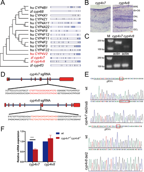
Generation of the cyp4v7 and cyp4v8 double knockout zebrafish. (A) Phylogenetic relationships and gene structures of CYP4 family genes in human and zebrafish. (B) In situ hybridization of cyp4v7 and cyp4v8 in retinas of two-month-old WT zebrafish. IS+OS, inner segment and outer segment; ONL, outer nuclear layer; INL, inner nuclear layer; GCL, ganglion cell layer. Scale bars: 20 µm. (C) Semiquantitative RT-PCR analysis of cyp4v7 and cyp4v8 expression in two-month-old zebrafish eyes. (D) CRISPR/Cas9 target sites. (E) DNA sequencing of the corresponding genomic region for WT and mutant zebrafish. The sequencing traces revealed a 2 bp deletion/6 bp insertion mutation in cyp4v7 and a 2 bp deletion mutation in cyp4v8. (F) Relative mRNA levels of cyp4v7 and cyp4v8 were detected by qPCR in WT and cyp4v7 and cyp4v8 double knockout zebrafish at 1 mpf. The results are shown as mean ± SD. ***P < 0.001.

Expression Data
Genes:
Fish:
Anatomical Terms:
Stage Range: Days 30-44 to Days 45-89

Expression Detail
Antibody Labeling
Phenotype Data
Fish:
Observed In:
Stage: Days 30-44

Phenotype Detail
Acknowledgments
This image is the copyrighted work of the attributed author or publisher, and ZFIN has permission only to display this image to its users. Additional permissions should be obtained from the applicable author or publisher of the image. Full text @ Invest. Ophthalmol. Vis. Sci.