
Figure 4
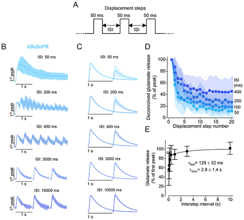
A, schematic representation of the stimulus protocol used to displace the cupula of the neuromasts towards the excitatory direction. Step displacements of 50 ms in duration, which saturated the fast glutamate response, were delivered with varying ISIs. B, average iGluSnFR responses to a train of steps with different ISIs. Traces are normalised to the peak response of the first step. Note that, for longer ISIs (3000 ms and 10 000 ms), image acquisition was interrupted in-between steps to limit photobleaching. C, deconvolved glutamate responses to paired pulses for different ISIs (see Methods). Individual traces were normalised to the amplitude of the response elicited by the first displacement step. In (B) and (C), continuous lines indicate the mean value and the shaded area indicates the SD. D, glutamate release (normalised peak) plotted as a function of pressure step number for the ISIs indicated on the right. E, time course of the recovery of the fast component of release. Peak glutamate release measured at the second displacement step as a function of ISI. The individual data points were fitted with a double exponential function: urn:x-wiley:00223751:media:tjp14725:tjp14725-math-0006, with urn:x-wiley:00223751:media:tjp14725:tjp14725-math-0007. Number of hair cells: 10 (50 and 100 ms), 11 (400 ms), 12 (200 ms, 1 and 10 s), 15 (3 s). Data are from 33 neuromasts (16 zebrafish). For the individual recordings used to calculate the averages shown in (D) and (E), see Supporting Information - Expanded dataset (Fig. 4). |