|
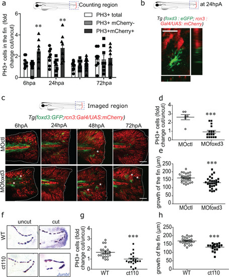
<italic>Foxd3</italic><sup><italic>+</italic></sup> NCdC are required for blastemal cell proliferation and zebrafish caudal fin fold regeneration.a Quantification of cell proliferation in the blastema of Tg(rcn3:Gal4/UAS:mCherry) larvae from 6hpA to 72 hpA. Mitotic cells were detected using an anti-PH3 antibody. Data are shown as fold change relative to the age-matched uninjured controls, and are the mean ± SEM, n = 8 (for the 6, 24, 72 hpA time points), n = 12 (for the 48 hpA time point), **p < 0.01. b Image of a time-lapse z-stack sequence of a Tg(foxd3:eGFP-F/rcn3:Gal4/UAS:mCherry) larvae at 24 hpA with x and y projections using the Fiji software (Scale bar = 8 µm). c Confocal images of caudal fin fold of Tg(foxd3:eGFP-F/rcn3:Gal4/UAS:mCherry) larvae injected with MOctl or MOfoxd3 at 3 dpf, and then from 6 hpA to 72 hpA (asterisks show pigments, scale bars = 80 µm, representative of n = 5 larvae from 3 independent experiments). d Blastemal cell proliferation in foxd3 and control morphants was assessed using an anti-PH3 antibody at 24 hpA. Data are shown as fold change relative to the age-matched uninjured controls, and error bars are the SEM, n = 6 (for the control group) and n = 16 (for the foxd3 morphants) biologically independent larvae, one-tailed Mann–Whitney test was performed, p = 0.0007, ***p < 0.001. e Quantification of caudal fin fold length in foxd3 and control morphants at 72hpA. Error bars are the SEM, n = 21 biologically independent larvae, one-tailed Mann-Whitney test was performed, p = 0.0002, ***p < 0.001. f The mRNA expression of the blastemal marker junb-l (blue) was detected by in situ hybridization at 24 hpA in control and amputated fin folds from 4 dpf wild type (WT) and Tg(foxd3:mCherry)ct110 (ct110) mutant larvae (representative of n = 15 biologically independent larvae per groups). g Blastemal cell proliferation in heterozygotes (WT) and homozygotes (ct110) Tg(foxd3:mCherry)ct110 larvae at 24 hpA was detected using an anti-PH3 antibody (graph represents the mean number of positive cells ± SEM, n = 21 (for the WT groups) and n = 18 (for mutants) larvae from 3 independent experiments, one-tailed Mann-Whitney test was performed, p = 0.0002, ***p < 0.001). (h) Quantification of fin fold growth at 72hpA in WT and Tg(foxd3:mCherry)ct110 mutant 6 dpf larvae (graph represents the mean ± SEM, n = 22 (for the WT group), n = 19 (for the mutant group) biologically independent larvae, one-tailed Mann Whitney test was performed, p = 0.0001, ***p < 0.001).
|