|
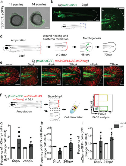
<italic>Foxd3</italic><sup>+</sup>NCdC in the developing and regenerating caudal fin fold mesenchyme are located next to proliferative <italic>rcn3</italic><sup>+</sup> mesenchymal cells.a Confocal images of Tg(foxd3:eGFP-F) zebrafish embryos at 11 somites and 14 somites (Scale bars = 300 µm). b, c Representative fluorescent images of Tg(foxd3:eGFP-F) 3 dpf larvae (Scale bar = 400 µm. (b) scale bar = 80 µm (c) d Schematic representation of caudal fin fold regeneration after amputation of 3 dpf larvae. e Representative confocal microscopy images of Tg(foxd3:eGFP-F/rcn3:Gal4/UAS:mCherry) larvae during regeneration (Scale bars = 80 µm, representative from 5 biologically independent larvae examined over 3 independent experiments). f Schematic representation of the fluorescent-activated flow cytometry analysis of eGFP+ and mCherry+ cells from caudal fin fold of Tg(foxd3:eGFP-F/rcn3:Gal4/UAS:mCherry) larvae at 6 and 24 hpA. Data were analysed using FlowJo v10. g Frequency of mCherry+ cell number in the caudal fin fold at 6 and 24 hpA relative to the age-matched uninjured controls. h Frequency of eGFP+ cell number in the caudal fin fold at 6 and 24 hpA relative to the age-matched uninjured controls (fold change). i Frequency of mCherry+eGFP+ cell number in the caudal fin fold at 6 and 24 hpA relative to the age-matched uninjured controls. g–i Graphs represent the mean value ± SEM, one-tailed Wilcoxon test was performed, *p < 0.05, p = 0.05 (g), p = 0,0143 (i), n = 50–300 larvae per group from 5 independent experiments.
|