|
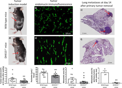
Tumor growth and metastases in DHX15<sup>+/−</sup> mouse.Macroscopic images of tumor size in wild-type (a) and DHX15+/- mice (d) three weeks after mouse Lewis lung cancer cells (LLC1) implantation. The arrows indicate the primary tumor. The quantification of tumor volume (cm3) is shown on the lower graph. **p < 0.01 vs. wild-type mice (n = 15 animals for each condition). Middle panels show endomucin immunostaining of intratumoral blood vessels in wild-type (b) and DHX15+/− mice (e). The quantification of the total vascular perimeter of all the intratumoral blood vessels that were positive for endomucin immunostaining was performed with the software Image J. Then, the total vascular perimeter per field was divided by the total number of endomucin+ vessels per field. The statistical comparison between experimental groups was made considering the result of this index (Perimeter/Vessel). **p < 0.01 vs. wild-type mice (n = 15 biologically independent samples for each condition; original magnification: ×200). Right panels show representative lung sections of lung metastatic area after haematoxylin-eosin staining (H&E) in wild-type (c) and DHX15+/− mice (f). The arrows indicate the metastatic areas. Quantifications of the percentage of lung metastases are shown in the graphs below. In the graph on the left: all tumors. *p < 0.05 vs. wild-type mice (n = 15 biologically independent samples for each condition). In the graph on the right: primary tumors with similar size. **p < 0.01 vs. wild-type mice (n = 3 biologically independent samples for each condition). Original magnification: ×10. All statistical analyses were performed using unpaired two-tailed Student’s t-test. All bar graphs are presented as mean ± SEM.
|