|
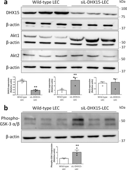
Characterization of the Akt and DHX15 signaling crosstalk in endothelial cells.a The expression of DHX15, Akt1, and Akt2 proteins was evaluated by western blot using cell lysates from wild-type and siL-DHX15-LECs. β-actin was used as a loading control. The densitometric analysis of the protein expression is shown on the bar graph. **p < 0.01 vs. wild-type LEC (n = 3 biologically independent samples for each condition). b The activity of Akt was determined in non-silenced and DHX15-silenced endothelial cells in a kinase reaction using recombinant GSK-3 as substrate. The levels of GSK-3α/β phosphorylation were analyzed by western blot using a phospho-GSK-3 specific antibody. β-actin was used as a loading control. The densitometric analysis of the protein expression is shown on the bar graph. **p < 0.01 vs. wild-type LEC (n = 3 biologically independent samples for each condition). All statistical analyses were performed using unpaired two-tailed Student’s t-test. All bar graphs are presented as mean ± SEM.
|