FIGURE

FIGURE 2

ID
ZDB-FIG-210817-24
Publication
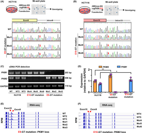
Lin et al., 2021 - Base editing-mediated perturbation of endogenous PKM1/2 splicing facilitates isoform-specific functional analysis in vitro and in vivo
Other Figures
All Figure Page
Back to All Figure Page
FIGURE 2

Splicing junction site mutations of PKM gene disrupt endogenous isoform‐specific gene splicing. a‐b. Schematic diagram of establishment of E9‐GT A, and E10‐GT B, mutant cell lines from single clones. The E9‐GT mutant cells were diluted and plated onto 96‐well plates for culture. After 10 d, the single clone‐derived cell lines were subjected to Sanger sequencing for genotyping. Three single clone‐derived cell lines were established for E9‐GT (Mut1‐3) and E10‐GT (Mut4‐6) mutant HCT116 cells. C, Detection of PKM1 and PKM2 expression by gel electrophoresis with expected length of PCR fragments in wild‐type (WT, WT2 and WT3), E9‐GT (Mut1‐3) and E10‐GT (Mut4‐6) mutant HCT116 cell lines. D, Semi‐quantitative analysis of PKM1 and PKM2 expression relative to GAPDH in c using ImageJ software. E‐F. Total RNA from wild‐type (WT, WT2 and WT3), E9‐GT (Mut1‐3) and E10‐GT (Mut4‐6) mutant HCT116 cell lines was subjected to RNA‐seq analysis. The enrichment of transcripts within exons or introns was presented by IGV viewer

Expression Data

Expression Detail
Antibody Labeling
Phenotype Data

Phenotype Detail
Acknowledgments
This image is the copyrighted work of the attributed author or publisher, and ZFIN has permission only to display this image to its users. Additional permissions should be obtained from the applicable author or publisher of the image. Full text @ Cell Prolif.