|
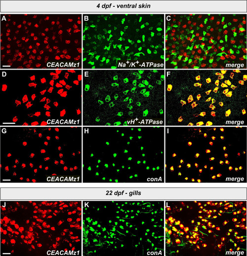
Identification of the ionocyte subtype that expresses CEACAMz1 on zebrafish epidermis.A-C. High magnification view of the yolk sac surface of a 4 dpf Tg(ceacamz1:mCherry-F) larva (A) immunolabelled with anti-Na+/K+-ATPase antibody (B); merge of A and B (C). D-F. High magnification view of the yolk sac surface of a 4 dpf Tg(ceacamz1:mCherry-F) larva (D) immunolabelled with anti-vH+-ATPase antibody (E); merge of D and E (F). G-I. High magnification view of the yolk sac surface of a 4 dpf Tg(ceacamz1:mCherry-F) larva (G) co-stained with Alexa Fluor 488-conjugated conA (H); merge of G and H (I). (J-L) High magnification view of the gill region of a 22 dpf Tg(ceacamz1:mCherry-F) larva (J) co-stained with Alexa Fluor 488-conjugated conA (K); merge of J and K (L). At all stages analyzed, CEACAMz1-expressing cells perfectly co-localize with cells labelled by the vH+-ATPase antibody or by conA. They do not overlap at all with the cells labelled by the Na+/K+-ATPase antibody.
|