|
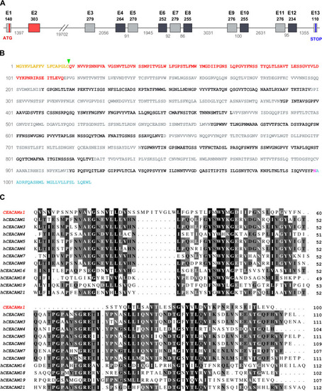
Sequence analysis of the <italic toggle='yes'>ceacamz1</italic> gene and its product.A. Organization of ceacamz1 coding region. The 35 kb-long ceacamz1 transcription unit is composed of 13 exons (red and grey boxes). The length in base pairs of the different exons and introns is indicated on the scheme. Each single Ig domain is encoded by a single exon (color-coded as in Fig 1). B. Protein sequence of CEACAMz1. The putative signal peptide is highlighted in orange and the green arrowhead indicates the most probable cleavage site (after Cys18). The Ig-V N-terminal domain is indicated in red. The Ig-C2 domains are highlighted in light grey (A-type) or dark grey (B-type). The ω-site for GPI-anchoring (Asn999) is displayed in purple. The residues highlighted in blue are cleaved off in the mature protein. C. Sequence alignment of the N-terminal Ig-V domain of CEACAMz1 with that of the twelve human CEACAMs. Amino acids are shaded from black to light grey according to their degree of conservation.
|