|
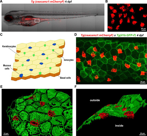
A. Live imaging of a 4 dpf Tg(ceacamz1:mCherry-F) zebrafish larva using spinning disk confocal microscopy. B. Zoom on the ventral region of the 4 dpf transgenic larva as indicated in panel A. C. Schematic representation of the cell composition of zebrafish skin epidermis at early larval stages. D. Z-projection of a portion of the ventral skin of a 4 dpf double-crossed Tg(ceacamz1:mCherry-F) x Tg(il1b:eGFP-F) zebrafish larva. CEACAMz1-expressing cells intercalate within the pavement of keratinocytes. E. and F. 3D-reconstructions of the Z-projection shown in panel D using IMARIS. Top view (E) and lateral view (F). CEACAMz1-expressing cells are embedded within the same layer as keratinocytes and they protrude on both sides of this layer.
|