|
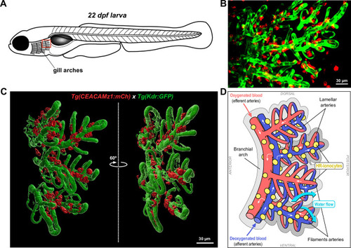
Distribution of CEACAMz1-positive ionocytes with respect to vasculature in the gills at late larval stages.A. Schematic representation of a juvenile zebrafish larva to highlight the part of the gills on which imaging was performed. B. Live imaging of a 22 dpf double-crossed Tg(ceacamz1:mCherry-F) x Tg(kdr:eGFP-F) larva using spinning-disk confocal microscopy. Z-projection centred on the most posterior arch of the gills. CEACAMz1-ionocytes (red) distribute along the branchial blood vessels (green), mainly the filament arteries at this stage. C. 3D reconstruction of the most posterior branchial arch using IMARIS software. The two distinct views are rotated by 60°. CEACAMz1-expressing cells are localized along the filament arteries on the inner side of the branchial arch, i.e. along the afferent arteries. D. Schematic representation of the 3D reconstruction visualized in C. Blood vessels are color-coded according to their oxygen content (efferent arteries in red, afferent arteries in blue). HR ionocytes expressing CEACAMz1 are displayed in yellow. The direction of water flow is indicated with blue arrows and is opposite to blood flow (white arrows).
|