|
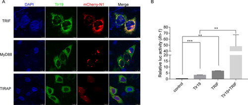
Tlr19 colocalizes and interacts with TRIF.A HeLa cells transiently transfected with Tlr19-GFP and TRIF-mCherry-N1, MyD88-mCherry-N1 or TIRAP-mCherry-N1. After 24 h, the cells were fixed with 4% paraformaldehyde, stained with DAPI and subjected to confocal microscopy analysis. Green signals represent CcTlr19, red signals display adaptors, and blue staining indicates the nucleus. B 293 T cells were transfected with empty vector, Tlr19, TRIF or Tlr19 + TRIF together with Luci-ifn and rhRL-TK reporter plasmids. After 48 h of transfection, all luciferase activities were calculated by normalization to Renilla luciferase activity, and the results are shown as the fold change compared to the control group (empty vector). Means ± SD (n = 3), **P < 0.01, ***P < 0.001, one-way ANOVA.
|