IMAGE

Figure 7

ID
ZDB-IMAGE-210707-29
Source
Figures for Shan et al., 2021
Image
Figure Caption

Figure 7

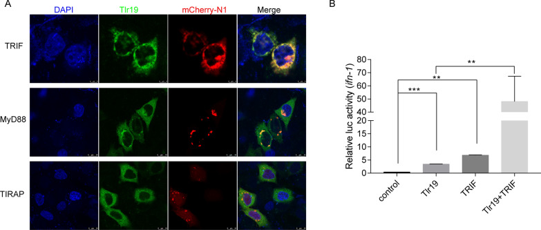
Tlr19 colocalizes and interacts with TRIF.A HeLa cells transiently transfected with Tlr19-GFP and TRIF-mCherry-N1, MyD88-mCherry-N1 or TIRAP-mCherry-N1. After 24 h, the cells were fixed with 4% paraformaldehyde, stained with DAPI and subjected to confocal microscopy analysis. Green signals represent CcTlr19, red signals display adaptors, and blue staining indicates the nucleus. B 293 T cells were transfected with empty vector, Tlr19, TRIF or Tlr19  +  TRIF together with Luci-ifn and rhRL-TK reporter plasmids. After 48 h of transfection, all luciferase activities were calculated by normalization to Renilla luciferase activity, and the results are shown as the fold change compared to the control group (empty vector). Means  ±  SD (n  =  3), **P  <  0.01, ***P  <  0.001, one-way ANOVA.

Acknowledgments
This image is the copyrighted work of the attributed author or publisher, and ZFIN has permission only to display this image to its users. Additional permissions should be obtained from the applicable author or publisher of the image. Full text @ Vet. Res.