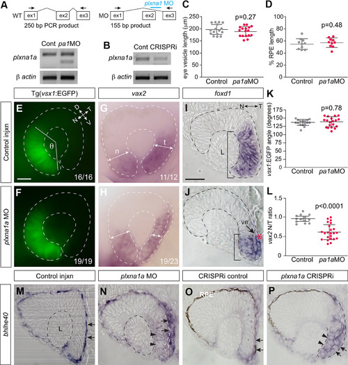
Plxna1a loss-of-function recapitulates the temporal eye defects observed in sema6d mutants. A, B, RT-PCR confirming morpholino mis-splicing (outlined in schematic) of plxna1a transcript (A), and knock down of plxna1a transcript in CRISPRi-injected embryos (B). β-Actin mRNA as loading control. C, D, Normal expansion of the bhlhe40-expressing RPE progenitor domain at the 14 ss. Mean anteroposterior eye vesicle length (C) and percent RPE expansion (D; RPE bhlhe40+ domain length/anteroposterior eye vesicle length) are not significantly different in plxna1a morphants as compared with controls [N = 2, control n = 17, plxna1 MO n = 17; p values are unpaired t tests, df = 33 (C), df = 18 (D), error bars are SD]. E, F, Tg(vsx1:GFP) expression in the nasal retina. G, H, vax2 mRNA in lateral views of a control (G) and a plxna1a morphant (H) 24-hpf eye. I, J, Transverse sections of whole-mount foxd1+ RNA ISH of 24-hpf eyes. Early ventral (future temporal) foxd1+ tissue undergoes rim movement into the outer leaflet in control (I), but in a plxna1a morphant remains partially in the inner leaflet (J; red asterisk and compare bars). Also evident is an open ventricle (arrow in J) in the morphant. K, The average angle formed by the lateral edges of the vsx1 (J) domain to the center of the lens (θ) is similar between controls and plxna1a morphants (N = 2; control n = 16, plxna1a MO n = 19, p value is unpaired t test, df = 33). L, Ratio of the width of the temporal to nasal (t and n in G) vax2 whole-mount RNA ISH domain measured in images of lateral eyes (unpaired t test, p < 0.0001, control n = 13, plxna1a MO n = 23, error bars are SD, df = 34). M–P, Transverse eye sections of whole-mount RNA ISH for bhlhe40+ performed on 24-hpf control embryos (M, O) or embryos injected at the one-cell stage with either an antisense plxna1a morpholino (N) or a sgRNA against exon5 of plxna1a along with dead-cas9 mRNA (CRISPRi; P). RPE bhlhe40+ progenitors elongate in control embryos (arrows in M, O) to line the back of the eye, and abut the lens, while the bhlhe40 signal is expressed ectopically in the morphant and CRISPRi-injected embryos (arrowheads N, P), and RPE cells retain a cuboidal shape (arrows N, P). Embryos in I, J, M, N were treated with 1-phenyl 2-thiourea to inhibit pigmentation of the RPE. Scale bars: 75 μm (E–H) and 50 μm (I, J, M–P). A: anterior, D: dorsal, L: lens, N: nasal, P: posterior, T: temporal, V: ventral, ve: ventricle.
|
