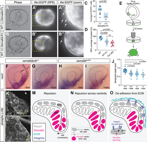
Possible Sema6d-Plxna1 repellent interactions during optic cup morphogenesis. A, B, EGFP+ eye vesicles from 18-ss wild-type Tg(tfec:EGFP) embryos were explanted and cultured in control media (A–A’’) or in the presence of a soluble Sema6d protein (Sema6d-Fc; B–B’’). A’’, B’’ are magnified views of the boxed areas in A’, B’. C, Quantitation of the average number of EGFP+ RPE cells that left the explant in the presence or absence of Sema6d-Fc (N = 3; n = 8–9 explants/condition each from a separate embryo, error bars are SD; unpaired t test, df = 16). D, Percent of GFP+ RPE coverage over the explanted Tg(tfec:EGFP) optic cup. Eye explants cultured in vitro develop RPE that covers the extent of the explant, whereas those cultured in the presence of soluble Semd6d fragment do not (N = 3; n = 8–9 explants/condition, each from a separate embryo, error bars are SD; one-way ANOVA, Dunnett’s multiple comparisons test, df = 24). E, Schematic of eye explant culture experiments. F–I, Lateral views of vax2 whole-mount ISH at 24 hpf. Losing Sema6d or inhibition of c-Abl with dasatinib disrupts temporal eye morphogenesis (asterisks). J, Quantitation of optic cup morphogenesis defects by representing the ratio of the width of the temporal versus nasal eye (N = 3; n = 14–18 embryos/condition, error bars are SD; one-way ANOVA, Dunnett’s multiple comparisons test, df = 59). K, L, Immunolabeling of cryostat sections through the eye vesicle of 18-hpf wild-type (K) and sema6d mutant (L) embryos for the phosphorylated form of c-Abl (N = 2 independent experiments). Dotted yellow lines indicate the separation between the neural retina and the RPE. Yellow arrowheads point to labeling of temporal eye vesicle in wild-type, with this label largely absent in mutants. M, Simple repulsion model. Our data support the possibility that Plxna1a from RPE cells activates Sema6d reverse signaling in neural retinal progenitors to promote movement of the cells of the inner eye vesicle around the distal rim of the optic cup. The possibility that Sema6d forward signals to Plxna1a-expressing RPE cells appears less likely. N, Repulsion across the ventricle model. Progenitor RPE cells interact with neural retina cells to allow leaflets to slide over each other during optic cup morphogenesis. Our data support the idea that Sema6d reverse signaling is the main mode involved. O, De-adhesion from ECM model. Progenitor RPE cells interact with neural retina cells to prevent over-adhesion to the ECM. Scale bars: 50 μm (A, F).
|
