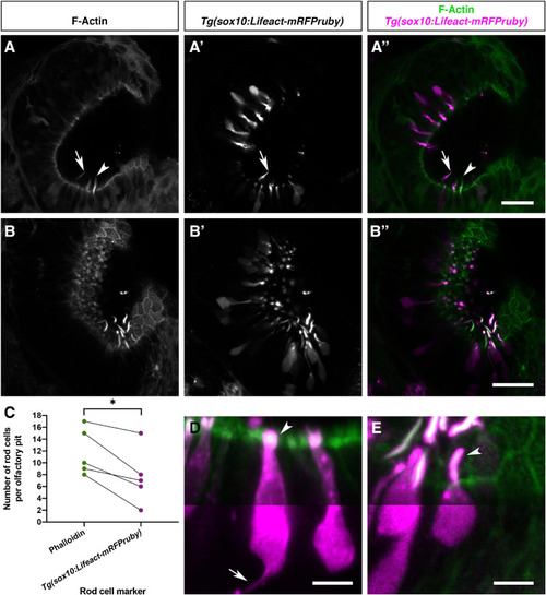
Olfactory rod cells are apically located in the zebrafish olfactory epithelium, with a rounded cell body and no detectable axon. (A–B″) Airyscan confocal image of Alexa-phalloidin signal (A,B), Tg(sox10:Lifeact-mRFPruby) signal (A′,B′), and merged signals (A″,B″) in olfactory pits of 4–5 dpf larvae; anterior to the top, lateral to the right. Arrowhead marks one olfactory rod negative for Lifeact-mRFPruby. Arrow marks one olfactory rod positive for Lifeact-mRFPruby. Scale bars = 20 μm. (C) Number of olfactory rod cells positively marked by Alexa-phalloidin (n of olfactory rods = 59), compared with the number of those also marked by Tg(sox10:Lifeact-mRFPruby) (n = 38), in olfactory pits of 4–5 dpf larvae (N of olfactory pits = 5). Connecting lines indicate olfactory rods from the same olfactory pit. Paired two-tailed t-test; * indicates P = 0.0146. (D) Enlargement of two microvillous OSNs, expressing Lifeact-mRFPruby, in the OE of a 4 dpf larva; Alexa-phalloidin signal (green), Tg(sox10:Lifeact-mRFPruby) signal (magenta). Arrowhead marks the microvillous apical projections. The gamma value for the magenta channel in the bottom half of the panel has been set to 0.5 to show the axon from one of the cells (arrow). Scale bar = 5 μm. (E) Enlargement of olfactory rod cells (of which both the apical actin projections and cell bodies are labelled by the Tg(sox10:Lifeact-mRFPruby) transgene) in the OE of a 4 dpf larva; Alexa-phalloidin signal (green), Tg(sox10:Lifeact-mRFPruby) signal (magenta). Arrowhead marks an olfactory rod cell apical projection, positive for both markers. The gamma value for the bottom half of the panel has been set to 0.5 as in panel (D); no axon is visible. Scale bar = 5 μm. See also Supplementary Movie 3.
|
