Figure S2
- ID
- ZDB-FIG-210219-13
- Publication
- Prentzell et al., 2021 - G3BPs tether the TSC complex to lysosomes and suppress mTORC1 signaling
- Other Figures
- All Figure Page
- Back to All Figure Page
|
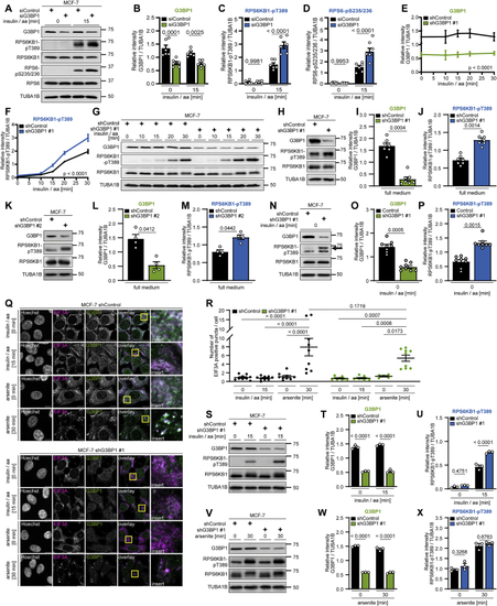
G3BP1 inhibits mTORC1 in cells without SGs, related to (A) Insulin/aa-stimulated siG3BP1 cells. n = 6. (B) Quantitation of G3BP1 in (A). Shown are data points and mean ± SEM. (C) Quantitation of RPS6KB1-pT389 in (A). Data shown as in (B). (D) Quantitation of RPS6-pS235/236 in (A). Data shown as in (B). (E) Quantitation of G3BP1 in (G). Mean ± SEM. (F) Quantitation of RPS6KB1-pT389 in (G). Data shown as in (E). (G) Time course analysis of shG3BP1 #1 cells, insulin/aa-stimulated for up to 30 min. n = 3. (H) shG3BP1 #1 cells cultured in full medium. n = 7. (I) Quantitation of G3BP1 in (H). Shown are data points and mean ± SEM. (J) Quantitation of RPS6KB1-pT389 in (H). Data shown as in (I). (K) shG3BP1 #2 cells cultured in full medium. n = 4. (L) Quantitation of G3BP1 in (K). Shown are data points and mean ± SEM. (M) Quantitation of RPS6KB1-pT389 in (K). Data shown as in (L). (N) Serum/aa-starved shG3BP1 #1 cells. Arrow, RPS6KB1-pT389 signal. n = 8, including re-analysis of improved contrast detections for data shown in (O) Quantitation of G3BP1 in (N). Shown are data points and mean ± SEM. n = 8, including re-analysis of data shown in (P) Quantitation of RPS6KB1-pT389 in (N). Data shown as in (O). n = 8, including re-analysis of improved contrast detections for data shown in (Q) IF of shG3BP1 #1 cells. Cells were either serum/aa-starved and stimulated with insulin/aa for 15 min; or serum-starved and treated with arsenite for 30 min. Overlay: white, EIF3A and G3BP1 co-localization; magenta, EIF3A; green, G3BP1; inserts, magnifications of yellow square. Scale bar, 10 μm. n = 3, except shG3BP1 #1, arsenite [0 min], n = 2. (R) Quantitation of data shown in (Q). Shown are data points and mean ± SEM. (S) Immunoblot performed in parallel to IF data in (Q). Insulin/aa-stimulated shG3BP1 #1 cells. n = 3. (T) Quantitation of G3BP1 in (S). Shown are data points and mean ± SEM. (U) Quantitation of RPS6KB1-pT389 in (S). Data shown as in (T). (V) Immunoblot performed in parallel to IF data in (Q). Arsenite-exposed shG3BP1 #1 cells. n = 3. (W) Quantitation of G3BP1 in (V). Shown are data points and mean ± SEM. (X) Quantitation of RPS6KB1-pT389 in (V). Data shown as in (W). |
Reprinted from Cell, 184(3), Prentzell, M.T., Rehbein, U., Cadena Sandoval, M., De Meulemeester, A.S., Baumeister, R., Brohée, L., Berdel, B., Bockwoldt, M., Carroll, B., Chowdhury, S.R., von Deimling, A., Demetriades, C., Figlia, G., Genomics England Research Consortium, de Araujo, M.E.G., Heberle, A.M., Heiland, I., Holzwarth, B., Huber, L.A., Jaworski, J., Kedra, M., Kern, K., Kopach, A., Korolchuk, V.I., van 't Land-Kuper, I., Macias, M., Nellist, M., Palm, W., Pusch, S., Ramos Pittol, J.M., Reil, M., Reintjes, A., Reuter, F., Sampson, J.R., Scheldeman, C., Siekierska, A., Stefan, E., Teleman, A.A., Thomas, L.E., Torres-Quesada, O., Trump, S., West, H.D., de Witte, P., Woltering, S., Yordanov, T.E., Zmorzynska, J., Opitz, C.A., Thedieck, K., G3BPs tether the TSC complex to lysosomes and suppress mTORC1 signaling, 655-674.e27, Copyright (2021) with permission from Elsevier. Full text @ Cell

