|
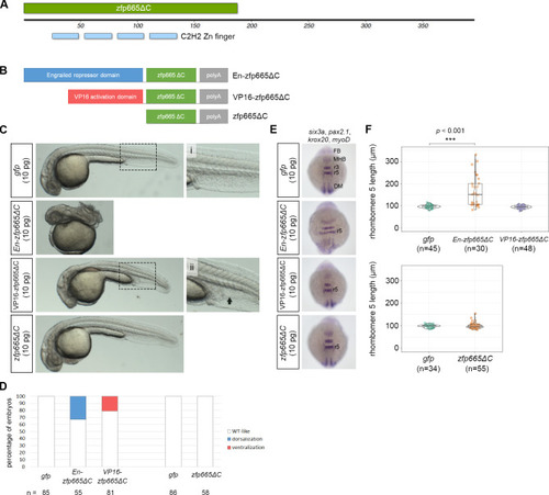
Functional validation of <italic>zfp665</italic> as a transcriptional repressor in zebrafish embryos.(A) A schematic representation of the protein domain organization of amphioxus zfp665, showing four consecutive C2H2-type zinc finger domains at the N-terminus. zfp665ΔC denotes the zinc finger region of zfp665 without its C-terminus. (B) A schematic representation of three different zfp665 fusion proteins, including engrailed repressor domain-zfp665ΔC fusion protein (En-zfp665ΔC), VP16 activation domain-zfp665ΔC (VP16-zfp665ΔC) fusion protein and the deletion of C-terminus of zfp665 (zfp665ΔC). (C) Phenotype of the zebrafish embryos injected with one of the three in vitro synthesized mRNA encoding the zfp665 fusion proteins at 24 hpf. (i) and (ii) are high-magnification views of the corresponding boxed areas. The black arrow indicates expanded ventral tissue. (D) Percentages of 24 hpf zebrafish embryos with the indicated phenotypes after injection of gfp (control) or one of the three in vitro synthesized amphioxus mRNAs encoding zfp665 fusion proteins. Only live larvae were scored. (E) WMISH of pax2.1, krox20, myoD and six3a in gfp (control) or indicated amphioxus mRNA-injected zebrafish embryos at the 10 somite stage. (F) Statistical analysis of the lengths of rhombomere 5 at the 10 somite stage in zebrafish embryos injected with gfp or one of the three forms of zfp665 mRNAs. DM, dorsal mesoderm; FB, forebrain; MHB, midbrain-hindbrain boundary; r3, rhombomere 3; r5, rhombomere 5. Statistical significance was determined by Welch’s one-way ANOVA with Games-Howell post hoc test plus Bonferroni correction when the number of groups was greater than 2 or Student’s t-test when the number of groups was 2. ***p < 0.001. Underlying data are available in S1 Data.
|