
Figure 7—figure supplement 1.
- ID
- ZDB-FIG-210113-144
- Publication
- Wague et al., 2020 - Mechanistic insights into volatile anesthetic modulation of K2P channels
- Other Figures
- All Figure Page
- Back to All Figure Page
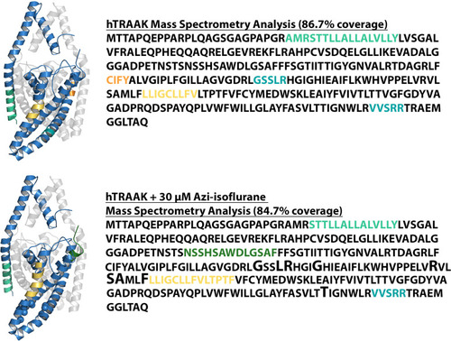
Results of MS analysis of TRAAK in the absence of reaction with azi-isoflurane (top) or following reaction with 30 μM azi-isoflurane (bottom). Regions positively identified by MS analysis are shown in blue in the TRAAK structural model (PDB ID 4WFE) and in black font in the sequence data. Regions absent from MS data are displayed in matching color in both the structural model and the sequence data. TRAAK residues homologous to TREK1 positions that exhibit high isoflurane occupancy in MD simulation are displayed as spheres in the structural model of TRAAK and are denoted in the sequence data by an enlarged font. All these residues are identified by MS, but none show evidence of azi-isoflurane labeling. A group of residues in the C-terminal region of TRAAK (marked in gray in the sequence data) were absent from our MS analysis but are not present in the TRAAK structural model. |